Unknown's avatar

MET Atlantoaxial (C1-C2)

Diagnosis

Position:  Atlas rotated right.

Motion Restriction:  Atlas resists left rotation on axis.

Patient is supine on the table with the operator sitting or standing at the head.

Operator grasps the head with the palms of the hands and flexes the head to approximately 30-45˚.MET C1-C2 Atlas R right 2


Operator introduces left rotation against the restricted barrier.MET C1-C2 Atlas R right 3

Patient is instructed to turn the head to the right against the operator’s resisting right hand with a light isometric contraction.

Following a 3-5 second contraction and subsequent relaxation, the operator increases left rotation to the next resistive barrier.

Repeat the right rotational effort against resistance 3-5 times.  Retest.

Unknown's avatar

Occipitoatlantal (C0-C1) Extension Restriction

Patient is supine on the table with the operator sitting at the head of the table.

Operator’s hands grasp the sides of the patient’s head with the lateral aspect of the index fingers monitoring along the posterior arch of the atlas and the rest of each hand controlling the head.

Operator introduces backward bending to the first barrier by rolling the head posterior around an axis of rotation through the external auditory meatus.OASupER3


Using the hands, the operator introduces translation of the head from right to left, maintaining the eyes parallel to the head of the table sensing for OASupER4resistance to movement at his index fingers. If resistance is felt, motion restriction is to backward bending, right side-bending, and left rotation (C0 flexed [F], side-bent left [Sleft], and rotated [Rright].  Something interfered with the right condyle gliding forward.

Using the hands, the operator introduces translation of the head from left to right sensing for resistance to movement at his index fingers.  If resistance is OASupER5encountered, the restriction is to backward bending, left side-bending, and right rotation (FSrightRleft).  Something interfered with the left condyle gliding forward.

Unknown's avatar

Cervical Spine Dx/Tx: Supine

A.  In the absence of dysfunction, side-bending of the typical cervical spine to the right allows the right facet to close and the left facet to open. Side-bending to the left allows the left facet to close and the right facet to open.

In determining if a facet can close, one would bias the paired facets into extension by translating the vertebral segment anteriorly.  From there, translating that segment from right to left tests the right side’s ability to close, while translating from left to right tests the left side’s ability to close. 

In determining if a facet can open, one would bias the paired facets into flexion by translating the vertebral segment posteriorly.  From there, translating that segment from right to left tests the left side’s ability to open, while translating that segment from left to right tests the right side’s ability to open.


B.  In the absence of dysfunction, side-bending the occiput to the right (right to left translation) onto the superior articular facet of C1 causes the right condyle to glide anteriorly (extend) on C1 and the left condyle to glide posteriorly (flex) on C1. 

In determining if the condyles can symmetrically glide anterior or extend, one would bias the occiput into extension by translating the condyles anteriorly on the superior articular facets of C1 (backward bending).  From there, side-bending the occiput to the right further tests the right side’s ability to extend.  Side-bending the occiput to the left (left to right translation) further test the left side’s ability to extend.

In determining if the condyles can symmetrically glide posterior or flex, one would bias the occiput into flexion by translating the condyles posteriorly on the superior articular facets of C1 (forward bending).  From there, side-bending the occiput to the right further tests the left side’s ability to flex; side-bending to the left further tests the right side’s ability to flex.


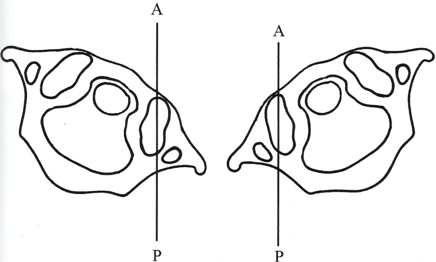
C.  The articular structures of C0-C1 are unique.  Relative to the anterior/posterior (sagittal plane), the anterior aspect of the articulating surface sits 30˚ medial to the posterior aspect of the articular surface.  Turning the occiput and C1 (atlas) to the right 30˚ places the right C0-C1 articular surface parallel to the A/P (anterior/posterior) plane.  Turning the occiput and C1 to the left 30˚ places the left articular surface parallel to the A/P plane.
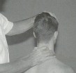
atlas


Unknown's avatar

Sitting Dx: Occipitoatlantal (C0-C1)

Patient sits on the table.

Operator stands behind with the thumb and index finger of the right hand grasping the posterior arch of the atlas and the left hand on top of the head to introduce movement.

Operator’s left hand introduces backward-bending, left side-bending, and right OAS3
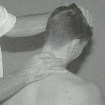
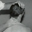
rotation of the head sensing for prominent fullness under the right thumb indicating posterior rotation of the atlas on the left.


Operator’s left hand introduces backward-bending, right side-bending, OAS4and left rotation of the head and monitors for prominence of the right posterior arch of the atlas under the right index finger, indicative of right rotation of the atlas.


Operator introduces forward-bending, right side-bending, and left OAS5
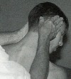
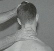
rotation with the left hand while monitoring for prominence of the right posterior arch of the atlas under the right index finger, indicating atlas right rotation.


Operator introduces forward-bending, left side-bending, and right OAS6rotation of the head with the left hand while monitoring for prominence of the left posterior arch of the atlas under the right thumb, indicating left rotation of the atlas.
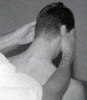
Unknown's avatar

Sitting Dx: Atlantoaxial (C1-C2)

Patient sits on the table with the operator behind.

Operator’s hands grasp the patients head and introduce forward bending to reduce rotation in lower typical vertebrae.AAS2


Operator introduces right rotation sensing for resistance to movement.AAS3


Operator introduces left rotation sensing for resistance to movement.AAS4


Unknown's avatar

Subscapularis Tendinosis versus Bicipital and Coracobrachialis Tendinosis

These conditions are typically called tendinitis.  A better term for both of these conditions might be tendon pain or tendinosis, which is the tearing of tendon fibers, due to repeated stress or overload, in the absence of an inflammatory process.  It is important to perform muscle resistance testing to determine which of these muscles are causing anterior shoulder pain.

The long head of the biceps tendon and the subscapularis tendon lie next to each other and general palpation may not be able to determine which muscle is creating the tendon pain.  Even though the pain may be in the same area, the treatment protocol and subsequent stretching are drastically different.

Subscapularis Treatment:subscap2-web

  • Follow the shoulder protocol starting with the velvet glove technique.
  • You will need to release all the other anterior shoulder muscles—pectoralis major, minor, and subclavius—before you work this muscle.
  • Next, follow the specific protocol for the subscapularis including multi-directional friction and eccentric contraction.
  • This is an extremely rare injury, as this muscle has a broad, flat, wide tendon, that is very deep and well protected. Although this muscle is almost always tight, subscapularis tendinosis is rare.

Biceps and Coracobrachialis Treatment:

  • Start with the basic shoulder protocol and release all of the anterior shoulder muscles.
  • The client is supine with the arm supinated, with support under the elbow using a bolster or a towel. Have the client perform a resisted test with the shoulder flexed to 90˚, palm facing up, to pinpoint the injury.
  • Follow the rest of the specific biceps/coracobrachialis protocol.
  • Continue to reassess and repeat any or all of the protocol until the client is pain-free.

biceps_coraco

Unknown's avatar

Sitting Dx: Typical Vertebrae (C3-C7)

Patient sits on table or treatment stool.

Operator stands behind with the thumb and index finger contacting zygopophysial joints bilaterally with the left hand on the vertex of the head to control motion.TCS2


Operator’s left hand introduces forward-bending, right side-bending, and right rotation while monitoring left zygopophysial joint for opening movement.TCS3


Operator introduces forward-bending, left side-bending, and left rotation, palpating for opening of the right zygopophysial joint by the operator’s right index fingerTCS4


Operator’s left hand introduces backward-bending, right side-bending, and right rotation with the right index finger monitoring the right zygopophysial joint’s capacity to close.TCS5


Operator introduces backward-bending, left side-bending, and left rotation with the left hand while monitoring the left zygopophysial joint’s capacity to close with the right TCS6thumb.

Unknown's avatar

Structural Diagnosis

The anatomy and biomechanics of the cervical spine result in five somatic dysfunctions.  The typical cervical segments (C3-C7) have non-neutral dysfunction with either a forward-bending or backward-bending restriction together with a coupled side-bending and rotation restriction to the same side (Type II motion).

At the atlantoaxial (C1-C2) junction, the primary somatic dysfunction is that of restriction of rotation to one side or the other.  While there may be minor forward/backward-bending and side-bending components to the rotational restriction, adequate treatment to the rotational restriction restores the minor movement motion simultaneously.

At the occipitoatlantal (C0-C1) junction, there are two dysfunctions possible.  There will either be forward-bending or backward-bending restriction with coupled side-bending and rotation to opposite side (Type I motion).

The structural diagnostic process starts by identifying levels of palpable deep muscle hypertonicity.  This identifies segments that need motion testing.  The diagnostic and the therapeutic processes seem to be most satisfactory by beginning from below and moving cephalad.

The bony landmark of most value in the typical cervical segment is the articular pillar.  They are palpated in the deep fascial groove between the semispinalis medially and the cervical longissimus laterally.  The paired examiner’s fingers can localize to the right and left articular pillars of any given cervical segment and introduce motion testing.

The typical cervical segment pillar is the size of the examiner’s finger pad.  The identification of the articular pillars begins by first identifying the spinous process of C2 and C7.  The C2 spinous process is the first bony prominence in the midline caudad to the external occipital protuberance (inion).  The spinous process of C7 (vertebra prominens) is the spinous process that remains palpable during cervical backward-bending.  The articular pillars of C2 and C7 are at the same level as the spinous processes.  Placing the examiner’s fingers between the pillars of C2 and C7 puts the finger pads in contact with with C3, C4, C5, and C6.  This provides the ability to localize to any specific cervical segment.  The structural diagnostic process can be performed in patients in both sitting and supine positions.

Unknown's avatar

Ideal Alignment of the Cervical Region

Ideal alignment of the cervical region allows the head to be positioned with minimal muscular effort.  Ideal alignment is an inward lordotic curve with both the upper and lower cervical region in a position of slight extension.

The most common alignment impairment observed in the cervical spine is a forward head posture.  The forward head posture is characterized by a forward translation of the lower cervical region, and hyperextension of the cervical region with typicaly, an increased kyphotic curve in the thoracic region.

Normal                                             Forward Head

scan0003 - Copy - Copy                        scan0003 - Copy

scan0003                       scan0003 - Copy (2)

The muscular adaptations associated with a forward head position are shortening of the cervical spine extensors and a lengthening of the intrinsic cervical spine flexors.  The forward head position also requires increased activity of the extensor muscles of the cervical spine to counter balance the head against the effect of gravity.  The muscular adaptations that occur with a forward head position results in an increase in compressive forces acting on the articular facets.

Additional alignment faults may include an increase in the degree of upper cervical extension in comparison to that of the lower cervical spine, suggesting possible muscular adaptations in the suboccipital region.  These adaptations can include shortness of the suboccipital extensors, superior obliques, inferior obliques, and rectus capitus and lengthened position of the suboccipital flexors, rectus capitus lateralis and anterior muscles.