Unknown's avatar

Positional Release (PR)

Excerpted from:  (Chaitow, Leon.  Muscle Energy Techniques.  2006, Elsevier Limited.)

It is probable that the very first modern positional release technique was discovered by Lawrence Jones, DO, in 1984. He called this technique ‘strain/counterstrain’ (SCS).

Arthur Pauls (1992) accurately termed this counterstrain inspired orthobionomy technique ‘the homeopathy of bodywork’ because positional release techniques subtly allow the body to correct and heal itself.  As such, they are often superior to direct forms of bodywork which forcibly attempt to impose solutions upon dysfunctional structures.

Chaitow’s genius was synthesis, integrating ideas of different thinkers and clinicians, including DOs, DCs, PTs, MDs, and Bodyworkers.

Chaitow described and demonstrated accurate diagnostic and powerful treatment techniques.  In fact, counterstrain diagnostics have been found to be more accurate than standard osteopathic and chiropractic assessments of patients with neck pain (McPartland, Goodridge & Brodeur, 1996).  In this study, the strain/counterstrain method of diagnosis known as ‘provacative palpation’ was found to be superior to three traditional palpation methods:  assessment of segmental motion (ROM range of motion), local tissue texture changes, and elicitation of pain of joint capsule tenderness.

Patients prefer positional release treatments: there are fewer side effects and the methods are easier to learn.  As John Goodridge, DO has suggested, ‘positional release techniques are the future’.

Several different methods exist that involve to positioning of an area of the body, or the whole body, in such a way as to evoke a therapeutically significant physiological response which helps to resolve musculoskeletal dysfunction.  The means whereby these beneficial changes occur seem to involve a combination of the circulatory and neurological changes that take place when a distressed area is placed in its most comfortable, its most ‘easy’, most pain free position.  The developer of the positional release technique known as ‘functional technique’, Harold V. Hoover (1969), termed this position ‘dynamic neutral’.  Charles Bowles (1969) has further discussed dynamic neutral:

Dynamic neutral is the state in which tissues find themselves when the motion of the structure they serve is free, unrestricted and within the range of normal physiological limits… Dynamic neutral is not a static condition… it is a continuing state of normal, living motion, during living activity… it is the state and condition to be restored to a dysfunctional area.”

The terms ‘ease’ and ‘bind’ are frequently used to describe the extremes of restriction and freedom of movement.  The term ‘dynamic neutral’ is considered interchangeable with ‘ease’.

The position of ‘ease’ that Jones discovered was an exaggeration of the position in which spasm was holding his patient.  Since this discovery, that a position which exaggerated a patient’s distortion could provide an opportunity for a release of spasm and hypertonicity, several variations on this basic theme have emerged.

The commonality of all these approaches is that they move the patient or the affected tissues away from any resistance barriers and toward positions of comfort… the position of ‘ease’.  Some approaches require verbal feedback from the patient regarding the degree of tenderness in a ‘tender point’ which is being used as a monitor, and which the operator is palpating while attempting to find a position of ease.  Other ‘functional’ methods or ‘positional release’ approaches involve the operator finding the position of maximum ease by means of palpation alone.

It is important to note that if positional release methods are being applied to chronically fibrosed tissue the result would produce a reduction in hypertonicity but would not result in any reduction of fibrosis.  Pain relief or improved mobility may be only temporary or partial in such cases.

Unknown's avatar

MET Occipitoatlantal (C0-C1) Condylar Glide

Diagnosis

  • Position:  ESrightRleft
  • Motion Restriction:  Forward-bending, left side-bending, and right rotation.

Treatment

Patient is supine on the table.

Operator’s left thumb and index finger grasp the posterior arch of the atlas and the occiput lies in the palm of the operator’s left hand.  Operator’s right hand spans the frontal region of the patient’s head.CG2


Operator rolls the head into forward-bending and with the right hand introduces left side-bending and right rotation against the resistance of the atlas CG4held by the operator’s left hand.

A 3-5 second mild isometric contraction of the head into backward-bending, or right side-bending is resisted by the operator’s hands.

After relaxation, the new barriers are engaged and the patient repeats the isometric contractions 3-5 times.

Retest.

Diagnosis

  • Position:  FSleftRright
  • Motion Restriction:  Backward-bending, right side-bending, and left rotation.

Treatment

Patient and operator (hand) positions are the same as above.

Operator introduces backward-bending through rotation, side-bending to the right, and rotation to the left with the right hand. CG8

Patient provides mild isometric contraction in the direction of forward bending or left side-bending against resistance offered by the operator’s right hand and against the atlas stabilized by the operator’s left hand.

After relaxation, the new backward-bending, right side-bending, left rotational barriers are engaged and the patient repeats the isometric contraction 3-5 times.

Retest.

Unknown's avatar

MET Occipitoatlantal (C0-C1)-ext.

Diagnosis

  • Position:  ESrightRleft
  • Motion Restriction:  Forward-bending, left side-bending, right rotation.

Treatment

Patient is supine on the table with the operator sitting or standing at the head.

Operator’s left hand controls the patient’s occiput with the web of the thumb and the index finger along the soft tissues of the suboccipital area.OAext2


The operator’s right hand cups the chin with the index finger in front and the middle finger below the tip of the ramus.  The operator’s right forearm is OAext3
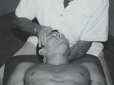
placed along the right side of the patient’s head.


Forward-bending is introduced by rotating the head forward by the operator’s hands around a transverse axis through the external auditory meatus.OAext4


Left side-bending and right rotation are introduced by the operator’s right forearm with slight left-to-right translation of the patient’s head to engage the restrictive barrier. OAext5(Right rotation is not actively introduced)


Patient instruction is to push the head directly posterior toward the table and into the hand offering resistance for 3-5 seconds of a mild isometric contraction. (An eye motion activating force is to look up toward the operator or toward the eyebrows.)

After relaxation, the operator engages the new forward-bending, left side-bending, and right rotational barriers.

Patient repeats the isometric contractions 3-5 times.

Retest.

Unknown's avatar

MET Occipitoatlantal (C0-C1)-flex.

 Diagnosis

  • Position:  FSrightRleft
  • Motion Restriction:  Backward-bending, left side-bending, right rotation.

Treatment

Patient is supine on the table with the operator sitting or standing at the head.

Operator’s left hand controls the patient’s occiput with the web of the thumb and the index finger along the soft tissues at the cervicocranial junction.OAflex2


Operator’s right hand holds the patient’s chin with the index finger in front and the middle finger below the tip of the ramus and with the right forearm in OAflex3contact with the right side of the patients head.


The backward-bending barrier is engaged by the operator’s hands rotating the head posteriorly around a transverse axis through the external auditory meatus.OAflex4


Left side-bending is introduced through the operator’s right forearm by slight left-to-right translation. (Rotation is not actively introduced)OAflex5


The patient is instructed to look down at the feet or pull the chin toward the chest against resistance offered by the operator’s right hand for a 3-5 second light isometric muscle contraction.

After relaxation, the new backward-bending, left side-bending , and right rotational barriers are engaged.

Operator’s muscle contraction is repeated 3-5 times with the operator relocalizing against the resisted barrier after each effort.

Unknown's avatar

MET Atlantoaxial (C1-C2)

Diagnosis

Position:  Atlas rotated right.

Motion Restriction:  Atlas resists left rotation on axis.

Patient is supine on the table with the operator sitting or standing at the head.

Operator grasps the head with the palms of the hands and flexes the head to approximately 30-45˚.MET C1-C2 Atlas R right 2


Operator introduces left rotation against the restricted barrier.MET C1-C2 Atlas R right 3

Patient is instructed to turn the head to the right against the operator’s resisting right hand with a light isometric contraction.

Following a 3-5 second contraction and subsequent relaxation, the operator increases left rotation to the next resistive barrier.

Repeat the right rotational effort against resistance 3-5 times.  Retest.