Unknown's avatar

Syracuse University Swim Study

Cycling and running require a certain amount of skill, but these skills are relatively simple compared with the technique required in swimming.  Because of the complexity of swimming and difficulties in research design, strength training to increase swimming speed may appear to be ineffective.  It has been postulated that the lack of a positive transfer between dry-land strength gains and swimming propulsive force may be due to the specificity requirements of swim training.  In other words, common gym-based training patterns may not be specific enough to actual pool-based swim patterns to result in a notable positive transfer.

Proper Periodization of Strength during the General Adaptation Phase and Maximal Strength Phase and improved Specificity of Training during the Conversion Phase would yield great improvements in swimming speed and endurance for all competitive swimmers, especially for females, juniors, and everyone at the masters level. Continue reading

Unknown's avatar

The Biomotor Abilities

Some confusion arises when endurance athletes and coaches question the need for the force produced in a maximal strength contraction.  Of course, a maximal level of force is not required to complete a stride, cycle revolution, or swim stroke.  So, why then is (maximal) strength considered a critical component for endurance athletes?  Because training maximal strength is the only way to develop synchronous motor unit recruitment, which is the primary requirement for increasing propulsive force and resisting muscular fatigue over long-endurance performances. Continue reading

Unknown's avatar

Dalton Demos

or choose

Unknown's avatar

Janda’s Classification of Muscle Imbalance Patterns

Excerpted from:  (Page, Frank, Lardner.  Assessment and Treatment of Muscle Imbalance, The Janda Approach.  2010, Human Kinetics, Champagne IL.)

Janda found that the typical muscle response to joint dysfunction is similar to muscle patterns found in upper motor neuron lesions, concluding that muscle imbalances are controlled by the CNS.  Janda believed that muscle tightness or spasticity is predominant.  Often weakness from muscle imbalance results from reciprocal inhibition of the tight antagonist.  The degree of tightness and weakness varies between individuals but the pattern rarely does.  These patterns lead to postural changes and joint dysfunction and degeneration.

Janda identified three stereotypical patterns associated with distinct chronic pain syndromes:  the upper crossed, lower crossed, and layer syndromes.  These syndromes are characterized by specific patterns of muscle tightness and weakness that cross between the dorsal and ventral sided of the body.

Upper-crossed syndrome (UCS) is also known as proximal or shoulder girdle crossed syndrome.

scan0002

The UCS pattern of imbalance creates joint dysfunction, particularly at the atlanto-occipital joint, C4-C5 segment, cervicothoracic joint, glenohumeral joint, and T4-T5 segment.  These focal areas of stress within the spine correspond to transitional zones in which neighboring vertebrae change in morphology.  Specific postural changes are seen in UCS, including forward head posture, increased cervical lordosis and thoracic kyphosis, elevated and protracted shoulders,and rotation or abduction and winging of the scapulae.  These postural changes decrease glenohumeral stability as the glenoid fossa becomes more vertical due to serratus anterior weakness leading to abduction, rotation, and winging of the scapulae.  This loss of stability requires the levator scapula and upper trapezius to increase activation to maintain glenohumeral centration.

Lower-crossed Syndrome (LCS) is also known as distal or pelvic crossed syndrome.

LCS

This pattern of imbalance creates joint dysfunction, particularly at the L4-L5 and L5-S1 segments, SI joint, and hip joint.  Specific postural changes seen in LCS include anterior pelvic tilt, increased lumbar lordosis, lateral lumbar shift, lateral leg rotation, and knee hyperextension.

If the lordosis is deep and short, then imbalance is probably in the pelvic muscles; if the lordosis is shallow and extends into the thoracic area, then imbalance predominates in the trunk muscles

Janda identified two subtypes of LCS.  Type A patients use more hip flexion and extension movement for mobility.  Their standing posture demonstrates an anterior pelvic tilt with slight hip flexion and knee flexion.  These individuals compensate with hyperlordosis limited to the lumbar spine and with a hyperkyphosis in the upper lumbar and thoracolumbar segments.

Jandas LCS Type B involves more movement of the low back and abdominal area.  There is minimal lumbar lordosis that extends into the thoracolumbar segmants, kyphosis in the thoracic area, and head protraction.  The COG is shifted backward with the shoulders behind the axis of the body, and the knees are in recurvatum.

Deep stabilizing muscles responsible for segmental spinal stability are inhibited and substituted for by activation of the superficial muscles.  Tight hamstrings may be compensating for anterior pelvic tilt or an inhibited gluteus maximus.

LCS also affects dynamic movement patterns.  If the hip loses its ability to extend in the terminal stance, there is a compensatory increase in anterior pelvic tilt and lumbar extension.  This compensation creates a chain reaction to maintain equilibrium, in which the increased pelvic tilt and anterior lordosis increase the thoracic kyphosis and cervical lordosis.

In adults, muscle balance begins distally in the pelvis and continues proximally to the shoulder and neck area.  In children, this progression is reversed, and muscle imbalance begins proximally and moves distally.

Janda’s Layer Syndrome (also referred to as stratification syndrome) is a combination of UCS and LCS.

Layer Syndrome

Patients display marked impairment of motor regulation that has increased over time and have a poorer prognosis than those with isolated UCS or LCS due to the long-standing dysfinction.  Layer syndrome is often seen in older adults and in patients who underwent unsuccessful surgery for herniated nuclus pulposus.

While chronic pain is difficult to treat, clinicians must be able to recognize Janda’s UCS, LCS, or Layer Syndrome in order to provide appropriate treatment.

Unknown's avatar

Causes of Muscle Tightness and Weakness

Excerpted from:  (Page, Frank, Lardner.  Assessment and Treatment of Muscle Imbalance, The Janda Approach.  2010, Human Kinetics, Champagne IL.)

Muscle tension (tone) is the force with which a muscle resists being lengthened.  Muscle tension may also relate to a muscle’s activation potential or excitability; thus, testing muscle tension has two components: viscoelastic and contractile.  The viscoelastic component relates to the extensibility of structures, while the contractile component relates to the neurological input.

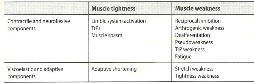
Contractile and Non-contractile Components of Muscle Tightness and Weakness

Causes tightnessWeakness

Muscle Tightness

Muscle tightness is the key factor in muscle imbalance.  Muscles prone to tightness are typically one third stronger than muscles prone to inhibition. Muscle tightness creates a cascade of events that lead to injury.  Tightness of a muscle reflexively inhibits its antagonist, creating muscle imbalance.  This muscle imbalance leads to joint dysfunction because of the unbalanced forces.  Joint dysfunction creates poor movement patterns and compensations, leading to early fatigue.  Overstress of activated muscles and poor stabilization lead to injury.

There are three important factors in muscle tightness:  muscle length, irritability threshold, and altered recruitment.  Muscles that are tight are usually shorter than normal and display an altered length-tension relationship.  Muscle tightness leads to a lowered activation threshold or lowered activation threshold, which means that the muscle is readily activated with movement.  Movement typically takes the path of least resistance, and so tight and facilitated muscles are the first to be recruited in movement patterns.  Tight muscles typically retain their strength, but in extreme cases they can weaken.

Structurally, increased muscle tension is caused by a lesion of the CNS that results in spasticity or rigidity.  Tight muscles are also described as hypertonic or facilitated.  Functionally, increased muscle tension results from either neuroflexive or adaptive conditions.  These two conditions are based on the contractile (neuroflexive factors) and viscoelastic (adaptive factors) components of muscle tension.

Neuroflexive Factors for Increased Tension

Limbic System Activation

Stress, fatigue, pain, and emotion contribute to muscle tightness through the limbic system.  Muscle spasms due to limbic system activation usually are not painful but are tender to palpation.  They are most frequently seen in the shoulders, neck, and low back and in tension headache.

Trigger Points (TrPs)

TrPs are focal areas of hypertonicity that are not painful during movement but are painful with palpation.  Essentially, they are localized, hyperirritable taut bands within muscle.

Muscle Spasm

Muscle spasm causes ischemia or an altered movement or joint position resulting from altered tension.  The spasm itself does not cause pain because spasm is not associated with increased EMG activity.  Muscle spasm is a typical response to joint dysfunction or pain irritation due to an impairment of interneuronal function at the spinal level.  Muscle spasm leads to a reflex arc of reciprocal inhibition for protection and subsequently impaired function of the motor system.  These muscles are also tender to palpation. 

Adaptive Factors for Increased Tension

Increased muscle tension also results from adaptive shortening.  Over time, muscle remains in a shortened position, causing a moderate decrease in muscle length and subsequent postural adaptation. Adaptive shortening is often considered overuse.  These muscles are not painful at rest but are tender to touch.  They exhibit a lowered irritability threshold and are readily activated with movement.  Over the long term, strength decreases as active fibers are replaced by non-contractile tissue.  It is thus extremely important for the clinician to identify the cause of increased muscle tension in order to apply the appropriate treatment.

Causes of Muscle Weakness

Muscle tension can decrease as a result of a structural lesion in the CNS such as a spinal cord injury or stroke.  A loss of tension leads to flaccidity or weakness.  Weak muscles are also describes as hypotonic or inhibited.  Functionally, muscle can be weak as a result of neuroflexive or adaptive changes and may exhibit delayed activation in movement patterns.

Neuroflexive Factors for Decreased Tension

Many contractile factors can contribute to decreased muscle tension.

Reciprocal Inhibition:  Muscle becomes inhibited reflexively when its antagonist is activated.  Weakness is often reflex-mediated inhibition secondary to increased tension of the antagonist.

Arthrogenic Weakness:  Muscle becomes inhibited via anterior horn cells due to joint swelling or dysfunction.  This weakness also leads to selective atrophy of Type II fibers.

Deafferentation:  Deafferentation is a decrease in afferent information from neuromuscular receptors.  Damage to joint mechanoreceptors (as seen with ligamentous injury) with subsequent loss of articular reflexes can cause altered motor programs, often influencing many muscles remote from the injured area.  This loss of afferent information ultimately leads to de-efferentation, or the loss of efferent signals to alpha motor neurons, which results in decreased muscle strength.

Pseudoperesis:  Pseudoperesis is a clinical presentation of neuroflexive origin.  Pseudoperesis has three clinical signs:  Hypotonia upon inspection and palpation, a score of 4/5 on a manual muscle test,and a change in the muscle activation pattern that may include delayed onset with early synergist activation or decreased EMG levels.  Faciliatory techniques often restore muscle strength and activation, but can also be inhibitory to a pseudoparetic muscle.

TrP Weakness:  Hyperirritable bands of muscle fiber decrease the stimulation threshold, leading to overuse, early fatigue, and ultimately weakness.  Muscles with active TrPs fatigue more rapidly than normal muscles and they exhibit a decreased number of firing motor units and poor synchronization.

Fatigue:  Muscle fatigue can be cause by metabolic or neurological factors.  Often during exercise muscles are fatigued before pain is experienced.  Thus the patient develops compensatory and faulty movement patterns before experiencing pain.

Adaptive Factors for Decreased Tension

Non-contractile factors causing decreased muscle tension are:

Stretch Weakness:  Stretch weakness is a condition in which muscle is elongated beyond physiological neutral but not beyond normal ROM.  Prolonged muscle elongation causes muscle spindle inhibition and the creation of additional sarcomeres.  The increased muscle length also changes the length-tension curve.  Stretch weakness is also known as positional weakness and is often associated with overuse and postural changes.  There is also an increase in the non-contractile tissue and a decrease in elasticity, leading to hypertrophy.  Ultimately overuse leads to ischemia and degeneration of muscle fibers, which further weakens the muscle.

When an inhibited and weak muscle is resisted, as is the aim of strengthening exercises, its activity tends to decrease rather than increase.  It is important to distinguish between neuroflexive weakness and structural weakness.  Often if the tight antagonist is stretched, the weak and inhibited muscle spontaneously increases in strength.

Unknown's avatar

Pathomechanics of Muscle Imbalance

Excerpted from:  (Page, Frank, Lardner.  Assessment and Treatment of Muscle Imbalance, The Janda Approach.  2010, Human Kinetics, Champagne IL.)

Muscles must be able to respond to a variety of simultaneous factors such as gravity, repetitive movement, and upright posture.  Muscles are influenced by both neurological reflexes and biomechanical demands, and therefore can be considered to be a window into the function of the sensorimotor system.  Janda found that patients with chronic musculoskeletal pain (most notably, chronic low back pain) exhibit the same patterns of muscle tightness and weakness as patients with CNS disorders exhibit.  This finding indicates a link between muscle imbalance and the CNS.

Tonic and Phasic Systems

The tonic system is the first used by the human body, as it is responsible for maintaining the fetal posture in newborn infants.  The phasic system soon is activated as the infant learns to lift her head for visual orientation.  The development of normal movement patterns utilizes reflexive coactivation of the tonic and phasic systems.  Muscles that are phylogenetically tonic demonstrate increased tone, while muscles that are phylogenetically phasic demonstrate decreased tone.  In patients with chronic musculoskeletal pain, this pattern of muscle imbalance manifests as tightness and weakness in the tonic and phasic muscles, respectively.  This finding supports Janda’s observation that chronic musculoskeletal pain is mediated by the CNS and is reflected in the sensorimotor system throughout the body.  It also allows us to predict typical muscle responses because of these neurodevelopmental chains.

Janda conceptualized muscle imbalance as an impaired relationship between muscles prone to tightness/shortness and muscles prone to inhibition.  More specifically, he believed that muscles predominantly static, tonic, or postural in function have a tendency to get tight and are readily activated in various movements—more so than muscles that are predominantly dynamic and phasic, which have a tendency to grow weak.

Tonic and Phasic Muscle systems:

scanTonicPhasic

Janda believed that muscles should not be classified based on the two-leg stance.  He preferred to consider the function of the muscle in relation to a one-leg stance, noting that muscles involved in maintaining upright posture during a single-leg stance (balancing) show a tendency toward tightness.

No muscle is exclusively tonic or phasic; some muscles may exhibit both characteristics.  Muscles do however have a tendency to be tight or weak in dysfunction.

Janda’s Classification of Muscles Prone to Tightness or Weakness

MusclesTightWeak

Czech physiotherapist Pavel Kolar (Kolar, 2001) expanded on Janda’s original list of tonic and phasic muscles from a more neurodevelopmental perspective adding to the tonic muscle list the coracobrachialis, brachioradialis, subscapularis, and teres major; and phasic muscle list the   rectus capitus anterior, suprspinatus, infraspinatus, teres minor, and deltoid.

Kolar also noted that the latissimus dorsi may be either tonic or phasic.  In contrast to Janda, Kolar categorized the piriformis and gastrocnemius as phasic muscles and suggested that the biceps, triceps and hip adductors exhibit both tonic and phasic portions.  Specifically, the long head of the triceps and short head of the biceps are tonic, while the medial and lateral triceps and long head of the biceps are phasic.  The short adductors are tonic, while the long adductors are phasic.

Faulty Movement Patterns

Janda theorized the facilitation of antagonists (flexors) and inhibition of agonists (extensors) in response to pain.  The subsequent muscle imbalances lead to changes in movement patterns.  Altered recruitment patterns typically begin with the delayed activation of a primary mover or stabilizer, along with early facilitation of a synergist.  Muscle tightness leads to overactivation of certain muscles, while muscles that should be activated are not, possibly due to inhibition or motor reprogramming.  Janda noted that altered peripheral input due to pain leads to these changes in muscle activation, causing faulty movement patterns that eventually become centralized in the motor program.

Imbalances in children begin in the upper extremity as opposed to the lower extremity, as is seen in adults.  These patterns of muscle imbalance are systematic and predictable because of the innate function of the sensorimotor system.  Subsequently, adaptive changes within the sensorimotor system (either vertical or horizontal) affect the entire system, most often progressing proximally to distally.  This muscular reaction is specific for each joint, suggesting a strong relationship between joint dysfunction and muscle imbalance.

Although Janda is considered the father of the neurological paradigm of imbalance, he recognized that muscle imbalances also occur as a result of biomechanical mechanisms.  Lifestyle often contributes to muscle imbalance as well.  Janda felt that the muscle imbalance in today’s society is compounded by stress, fatigue, and insufficient movement through regular physical activity as well as a lack of variety of movement.  This lack of variety contributes to repetitive movement disorders.  Janda noted that most repetitive movements reinforce the postural system, neglecting the phasic system, and lead to imbalance.

Unknown's avatar

Pathology of Musculoskeletal Pain

Excerpted from:  (Page, Frank, Lardner.  Assessment and Treatment of Muscle Imbalance, The Janda Approach.  2010, Human Kinetics, Champagne IL.)

Patients with chronic musculoskeletal pain continue to experience pain after a period of time that a peripheral pathology would normally resolve.  This persistent pain suggests a persistent  peripheral input.

These patients also exhibit altered pain processing in the CNS, as seen in the phenomenon of pain centralization.  Pain stimuli can alter sensitivity to central perception of pain and can alter the afferent signal at multiple levels, lowering pain thresholds in healthy regions throughout the body.  Thus clinicians should evaluate and treat chronic muscle imbalance and chronic musculoskeletal pain as a global sensorimotor dysfunction.

Janda believed that muscles, as opposed to bones, joints, and ligaments, are most often the cause of chronic pain.

Direct causes of muscle pain include muscle and connective tissue damage, muscle spasm and ischemia, and tender points or trigger points.  Most pain is associated with muscle spasm but is not the result of the spasm itself; rather the pain is caused by ischemia.

Indirect causes of muscle pain include altered joint forces due to muscle imbalance influencing movement patterns.  Joint dysfunction without spasm is typically painless.  Muscle imbalance can develop as a result of both acute and chronic pain.  Acute pain leads to a localized muscle response that changes the movement pattern to protect or compensate for an injured area.  Over time, this altered movement pattern becomes centralized in the CNS.  The viscious cycle pain and spasm has been questionable, but the viscious cycle of chronic pain involving the CNS and PNS seems plausible.

Components of the Chronic Musculoskeletal Pain Cycle:

Muscle Imbalance

Chronic pain is associated protective adaptive response in muscle in which agonists decrease in tone while antagonists increase in tone.  The pattern of neurological imbalance is based on neurodevelopment of the tonic and phasic muscle systems.  Muscle imbalance presenting with facilitation of an agonist inhibits the antagonist, possibly increasing the risk of injury.

Impaired Movement Patterns and Postural Changes

Postural responses to pain are common, facilitating the flexor response to protect the injured area.  The protective adaptation to pain through compensatory movement results in decreased range of motion and altered movement patterns.  Tightness of antagonists subsequently inhibits agonists based on Sherrington’s law of reciprocal inhibition.  This imbalance leads to further alterations in normal movement patterns.

Faulty Motor Programming and Motor Learning

Repetitive dysfunctional movement patterns eventually supersede the normal functional motor program because of the effect of motor learning.  The dysfunctional movement pattern becomes ingrained in the motor cortex as the new normal program for a specific movement pattern, thus reinforcing the dysfunctional pattern.

Altered Joint Forces and Altered Perception

Altered movement patterns change the normal patterns of joint stress.  Muscle imbalance alters joint position, changing the distribution of joint stresses on the joint capsule and surfaces.  Afferent input is essential in the modification of muscle activation to make movement well-coordinated and functional. Altered proprioception may be the actual cause of inhibition or spasm, not pain.

Joint Degeneration

Poor proprioception ultimately may be responsible for joint degeneration.  Muscle imbalance presents a much greater danger for joints than muscular weakness alone presents.  Therefore, functional pathology may in fact cause structural pathology.

Chronic Pain

Inflammatory mediators such as histamine and bradykinins are known to cause pain.  Joint pain and inflammation sensitize musculoskeletal afferent receptors.  Pain causes an adaptive response of muscle imbalance and altered posture and movement patterns and thus facilitates the vicious cycle.

Unknown's avatar

Pathomechanics of Musculoskeletal Pain and Muscle Imbalance

Vladimir Janda believed that pain is the only way the musculoskeltal system can protect itself.  Functional pathology of the sensorimotor system points to the importance of examining dysfunction rather than structural lesions.  Chronic musculoskeletal pain often arises from a functional pathology with resultant structural inflammation.  Structural lesions rarely cause pain themselves; rather, the inflammatory processes surrounding structural damage cause pain.  Often the site of pain is not the cause of pain; unfortunately, most clinicians focus on the area of chronic pain (structure) rather than the cause of pain (function).  An understanding of functional pathology forces clinicians to reevaluate their approach to the management of chronic musculoskeletal pain.

Excerpted from:  (Page, Frank, Lardner.  Assessment and Treatment of Muscle Imbalance, The Janda Approach.  2010, Human Kinetics, Champagne IL.)